Kidney Stones
 |
Kidney stones are calcifications in the urinary system that can cause pain, infection and, at times, can be dangerous enough to cause death. Kidney stones can be in any location in the urinary system.
Kidney Stones in the Kidney Proper
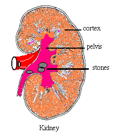
Here the stones are found mostly in the pelvis of the kidney. This is the major drainage part of the kidney. Sometimes, they can be in the Calyces, which are the small collecting parts in the kidney. If a kidney stone is in the pelvis of the kidney or the calyces of the kidney, depending on the size, shock wave treatment would be the best treatment. At times percutaneous lithotripsy is performed by putting a needle through the flank and then going in with wires inside the kidney. Of course, open kidney surgery is still available and is done for very large stones or stones which cannot be approached by the two above methods.
|
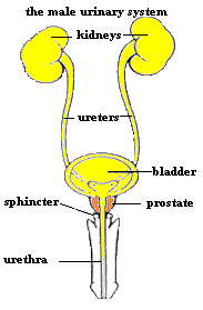 |
Stones in the Ureter
This is the duct that leads from the kidney to the bladder. If the stone is high in the ureter, it is best to push the stone back into the kidney and do ESWL (shock wave) on that. Sometimes, ESWL can be done with the stone in the upper ureter but success rates vary from 50% to 70%.Occasionally one has to remove the stones by ureteroscopy.
Mid-Ureter
If the stone is in the middle part of the ureter, we have to push it up into the kidney to do ESWL. In the middle part of the ureter, there is no good way to break up the stone. One can try ESWL but the success rate is 50%. Sometimes if there is blockage and fever, one has to go in and surgically remove it. Most often we place a stent to relieve pressure. Then either treat with ESWL or ureteroscopy. One has to wait one week for swelling to go down and the ureter to relax.
|
Stones in the Lower Part of the Ureter
Usually removed by a basket. This procedure consists of a basket that is packed inside a small catheter. The catheter is passed through a cystoscope inside the bladder and then up the ureter. It is inserted behind the stone so when it is opened, like an umbrella, it will catch the stone and bring it out. Sometimes it becomes necessary to dilate the ureter as in angioplasty to be able to insert a ureteroscope and remove the stone.Sometimes laser is used to break up the stone before one can safely remove it.
Bladder Stones
Bladder stones can usually be broken up by a machine called a Lithotriptor, or can be removed by surgery, depending on size.
Stones in the Prostate
Stones in the prostate are embedded in the substance of the prostate and cannot be removed or pulverized. They usually come out during TURP (transurethral removal of the prostate.)
Surgical Indications
- Fever - If fever develops during the course of the patients illness with a kidney stone or ureter stone, it means the urine behind the stone is turning into pus. If the pus gets into the blood stream then we have a condition called bacteremia. Bacteremia can be very dangerous and life threatening. So if fever develops, we usually operate on the patient within 24 to 48 hours.
- Pain - Some recurring pain is to be expected in the beginning. But if the pain is such that every three hours it is requiring intramuscular or IV injections, in about 48 hours we decide to intervene. Pain is not due to the stone but the blockage behind it.
- Obstruction - If the kidney is totally blocked and there is no pain or fever, treatment can wait a while. However, if treatment is delayed it can lead to kidney damage.
- Intervention - The pain due to a kidney stone is rarely ever due to the kidney stone itself. It is usually due to the distention or swelling of the kidney and the ureter behind the point of blockage. That is why sometimes, even if the stone is still retained, the pain may disappear for days because the urine is able to drain around the stone.
Management of Pain and Discomfort
- This consists of pain pills or shots.
- Fluids should be cut down in the beginning until the severe pain subsides. Then one can increase the fluids. Sometimes there is nausea, abdominal discomfort and bloating. These are all caused by the stone.
Prevention of Kidney Stones
After the stone is removed, it is sent for analysis.We will have an idea of what type of stone it is. This may not be enough, especially in patients with recurring episodes of kidney stones. One has to study the urine and the blood to see what was the cause of this specific type of stone formation. This is especially important, as there are multiple types of kidney stones. The most common is made of calcium and oxalate. The metabolic study of kidney stones consists of blood tests and 24 hour urine collection.
- On the first visit to the lab, the patient is instructed to collect 24 hour urine on a regular diet. This is done from 7:00 am to 7:00 am the next day, Then the urine is brought in and blood is obtained from the patient to compare the values.
- After two weeks, patient is seen in the office where results are discussed and he or she is placed on a special diet or medication, patient will be instructed to collect 24 hour urine to make sure the treatment is working. If not, the dose or the diet is adjusted. Repeating the work up in a year may be advisable. It is my impression that most patients who have done this metabolic work up have down very well and have not had any recurrent kidney stones.
- If the patient does not choose to have the work up, then a diet consisting of low salt, low oxalate, and low protein is advised. Increased fluids up to 60 ounces daily and increasing citrate, which is in orange juice and lemonade, have been known to prevent kidney stones.
|